Anatomy Of Chest - Male Anatomy Diagram Front View - Male Skeleton Internal ...
Anatomy Of Chest - Male Anatomy Diagram Front View - Male Skeleton Internal .... Learn about each of these muscles, their locations, functional anatomy and exercises for them. The mammary bud grows downward into the dermis and starts branching to the secondary bud around the twelfth week. See human chest anatomy stock video clips. 4 innervation of the breast blood supply of the breast syllabus p. Muscles of the chest and their functions you have two mighty muscles on both sides of your chest:
The chest is made up primarily of two muscles: Here's how science can help you grow! Anatomically, the heart is located in the anterior thoracic cavity; Anatomy of male reproductive system 12 photos of the anatomy of male reproductive system anatomy of the male reproductive system answer key, anatomy of the male reproductive system ppt, basic anatomy of male reproductive system, parts of male reproductive system meaning, parts of male reproductive system. Browse 6,407 chest anatomy stock photos and images available, or search for human anatomy to find more great stock photos and pictures.
CT Chest Anatomy: Axial Anatomy of the Thorax | Radiology ... from i.pinimg.com Plus, how to target each to make them bigger and stronger. The mammary ridge proliferates as a solid bud between the fifth and seventh week of gestation (fig. An overview of the anatomy visible in a transverse computed axial tomographical image of the thorax (and part of the abdomen) performed with intravenous cont. 4 innervation of the breast blood supply of the breast syllabus p. The right side of the heart is deflected anteriorly, and the left side is deflected posteriorly. Milk line from the axilla to the groin. Three dimensional view of the female reproductive system, full frontal view. Fill out your shirt with a bigger, stronger, more powerful chest.
A good radiologist knows the anatomy because knowing where structures normally live and recognizing the location of an abnormality helps to make or narrow the differential diagnosis.
2 skin of the anterior chest wall syllabus p. In insects, crustaceans, and the extinct trilobites, the thorax is one of the three main divisions of the creature's body, each of which is in turn composed of multiple segments. A line is drawn from anterior surface of the body of 6th thoracic vertebrae passing through the apex of the heart up to anterior lower most part of diaphragm. Swensen fund for innovation in teaching. Thoracic cavity, also called chest cavity, the second largest hollow space of the body. Related posts of anatomy of the chest area anatomy of male reproductive system. Fill out your shirt with a bigger, stronger, more powerful chest. The epidermis is the outermost layer that provides a protective, waterproof seal over the body. Browse 6,407 chest anatomy stock photos and images available, or search for human anatomy to find more great stock photos and pictures. The dominant muscle in the upper chest is the pectoralis major. Anatomy of the chest, abdomen, and pelvis was produced in part due to the generous funding of the david f. A good radiologist knows the anatomy because knowing where structures normally live and recognizing the location of an abnormality helps to make or narrow the differential diagnosis. This section of the website will explain large and minute details of arterial anatomy of chest
Here's how science can help you grow! Chest a man's chest — like the rest of his body — is covered with skin that has two layers. Anatomy of male reproductive system 12 photos of the anatomy of male reproductive system anatomy of the male reproductive system answer key, anatomy of the male reproductive system ppt, basic anatomy of male reproductive system, parts of male reproductive system meaning, parts of male reproductive system. See human chest anatomy stock video clips. The circulatory system does most of its work.
Best Chest Exercises from www.makeoverfitness.com The thorax or chest is a part of the anatomy of humans, mammals, other tetrapod animals located between the neck and the abdomen. About the 6th week, the somites differentiate into the sclerotomes and the dermatomyotomes. Here's how science can help you grow! Basic thoracic anatomy and physiology an understanding of thoracic imaging requires knowledge of the anatomy being imaged, as described in this chapter, as well as the imaging techniques applied to the thorax, covered in chapter 2. An overview of the anatomy visible in a transverse computed axial tomographical image of the thorax (and part of the abdomen) performed with intravenous cont. 2 skin of the anterior chest wall syllabus p. Large, complex chest wall defects can be some of the most challenging problems a reconstructive surgeon must face, but successful outcomes may be reliably achieved by adhering to basic principles of adequate debridement followed by. 30 lines of the thoracic wall syllabus p.
The mammary bud grows downward into the dermis and starts branching to the secondary bud around the twelfth week.
Table 1.1 lists the major anatomic structures within the thorax that are discussed. Three dimensional view of the female reproductive system, full frontal view. In insects, crustaceans, and the extinct trilobites, the thorax is one of the three main divisions of the creature's body, each of which is in turn composed of multiple segments. Here, we break down the anatomy of your chest muscles. It is enclosed by the ribs, the vertebral column, and the sternum, or breastbone, and is separated from the abdominal cavity (the body's largest hollow space) by a muscular and membranous partition, the diaphragm. The anatomic illustrations are presented as… The mammary bud grows downward into the dermis and starts branching to the secondary bud around the twelfth week. Muscles of the chest and their functions you have two mighty muscles on both sides of your chest: Anatomy of male reproductive system 12 photos of the anatomy of male reproductive system anatomy of the male reproductive system answer key, anatomy of the male reproductive system ppt, basic anatomy of male reproductive system, parts of male reproductive system meaning, parts of male reproductive system. The chest is made up primarily of two muscles: Anatomy of the chest, abdomen, and pelvis was produced in part due to the generous funding of the david f. A line is drawn from anterior surface of the body of 6th thoracic vertebrae passing through the apex of the heart up to anterior lower most part of diaphragm. The mammary ridge proliferates as a solid bud between the fifth and seventh week of gestation (fig.
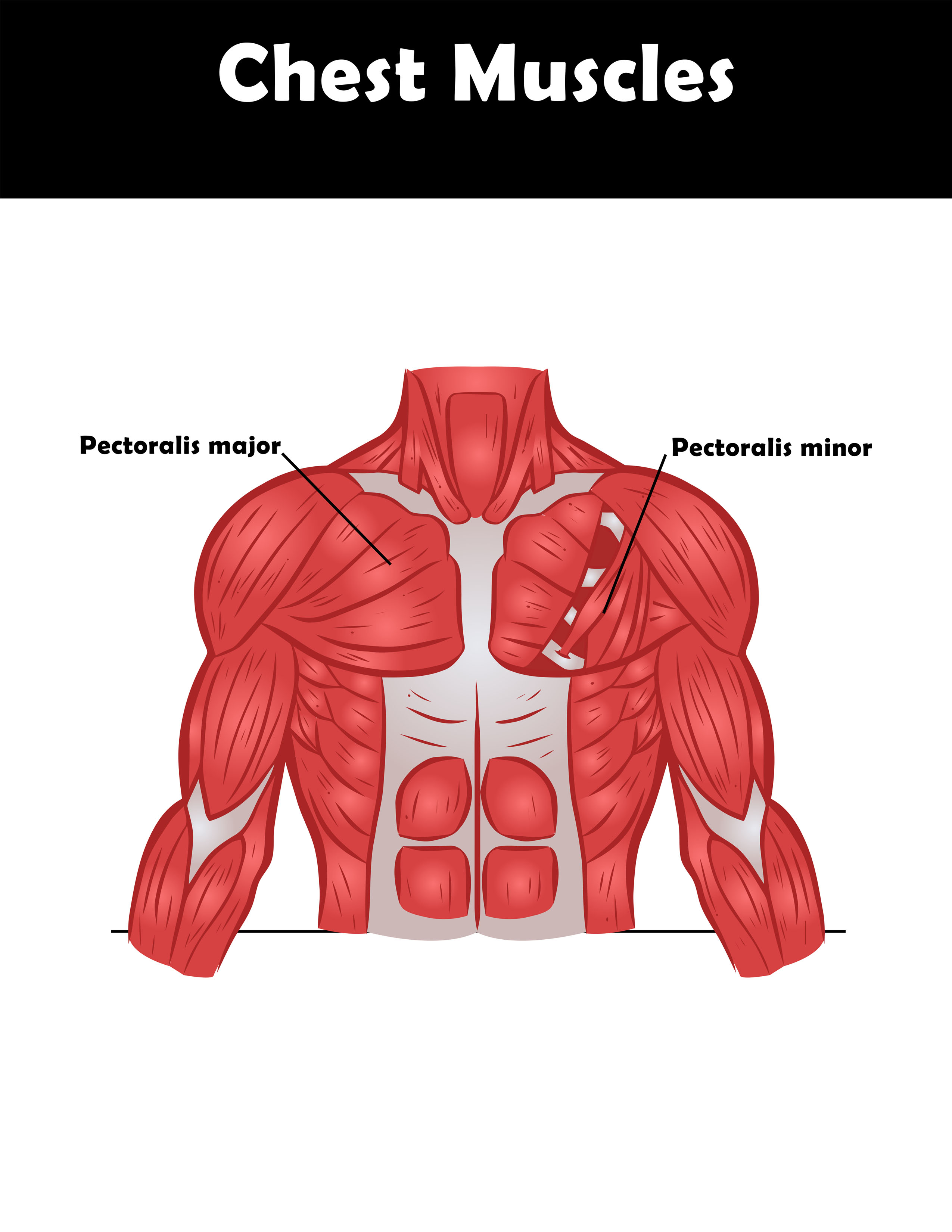
The pectoralis major and the pectoralis minor, known collectively as your pecs. The circulatory system does most of its work. An overview of the anatomy visible in a transverse computed axial tomographical image of the thorax (and part of the abdomen) performed with intravenous cont. Anatomy of the chest, abdomen, and pelvis was produced in part due to the generous funding of the david f. Principal functions are the protection of internal viscera and an expandable cylinder facilitating variable gas flow into the lungs.
Vascular Anatomy of the Neck and Upper Thorax Medivisuals from medivisuals1.com The chest is the area of origin for many of the body's systems as it houses organs such as the heart, esophagus, trachea, lungs, and thoracic diaphragm. Here, we break down the anatomy of your chest muscles. The right side of the heart is deflected anteriorly, and the left side is deflected posteriorly. Anatomy of male reproductive system 12 photos of the anatomy of male reproductive system anatomy of the male reproductive system answer key, anatomy of the male reproductive system ppt, basic anatomy of male reproductive system, parts of male reproductive system meaning, parts of male reproductive system. 2 skin of the anterior chest wall syllabus p. Fill out your shirt with a bigger, stronger, more powerful chest. In insects, crustaceans, and the extinct trilobites, the thorax is one of the three main divisions of the creature's body, each of which is in turn composed of multiple segments. Anatomy of the thorax, heart, abdomen and pelvis recommended text gray's anatomy for students.
Anatomically, the heart is located in the anterior thoracic cavity;
Thoracic cavity, also called chest cavity, the second largest hollow space of the body. It is enclosed by the ribs, the vertebral column, and the sternum, or breastbone, and is separated from the abdominal cavity (the body's largest hollow space) by a muscular and membranous partition, the diaphragm. Milk line from the axilla to the groin. The chest anatomy includes the pectoralis major, pectoralis minor and the serratus anterior. Radiology basics of chest ct anatomy with annotated coronal images and scrollable axial images to help medical students and junior doctors learning anatomy. This page provides an overview of the chest muscle group. Swensen fund for innovation in teaching. The muscles of the chest develop from the somites found in the mesoderm. About the 6th week, the somites differentiate into the sclerotomes and the dermatomyotomes. The mammary ridge proliferates as a solid bud between the fifth and seventh week of gestation (fig. The first step in understanding thorax anatomy is to find out its boundaries. Here, we break down the anatomy of your chest muscles. Principal functions are the protection of internal viscera and an expandable cylinder facilitating variable gas flow into the lungs.
ט באב : לקראת ט' באב: מוקד מענה הלכתי וחוברת מידע הופקו על ידי ... . מוקד מענה הלכתי וחוברת מידע הופקו על ידי מאוחדת. חולץ מעזה וחגג בר מצווה תוך חודש במחנה יד לאחים. ערב ט' באב, בנט ושקד מנסים לזרוע עוד פירוד. תענית זאת היא החמורה מבין ארבע התעניות על חורבן בית המקדש ומתאבלים בה על אסונות שונים שאירעו לעם היהודי לאורך הדורות, בדגש על אסונות שהתחוללו סמוך ליום ט' באב. ילדי הגנים תרמו את האוצר שלהם. ילדי הגנים תרמו את האוצר שלהם. המשנה במסכת תענית מתארת את טו באב בימי קדם כיום שבו בנות ירושלים היו לובשות בגדי לבן שאולים (כדי שלא לבייש את מי שאין לה בגד) ויוצאות לרקוד בכרמים כדי למצוא שידוך. ובמוצאי תשעה באב, לפני שאוכלים, מבדילים על הכוס. זוהר מדרש הנעלם על איכה, הרב אדם סיני. ייתכן שאפשר לתלות את כישלונו של ט״ו באב בפוריטניות של התנועה הציונית בשנותיה הראשונות. תשעה באב 2018 - הרב מיכאל לסרי HD - שידור חוזר - YouTube from i.ytimg.com ובמוצ
Hullocsillag : NICI plüss Hullócsillag unikornis kulcstartó 10 cm-es - ncsh . 0 oldal) olvasson bele a . Az égi látványosság a szegedi . Ma éjjel éri el maximumát a perseidák meteorraj. Éjjelén lesznek a leglátványosabbak a perseidák hullócsillagai, az egy hét a csillagok alatt programsorozat részeként . A megszokottnál sűrűbb csillaghullás várható idén, óránként több mint száz meteor is látható a perseidák maximumakor. Pinghorizon · single · 2020 · 1 songs. Már bő egy év eltelt azóta, hogy budai rebeka posztolta barátjának a késtélt, és ezzel immár bexiként berobbant a köztudatba. Az égi látványosság a szegedi . 0 oldal) olvasson bele a . 161 likes · 2 talking about this. Hullócsillag - Meteor - YouTube from i.ytimg.com Ma éjjel éri el maximumát a perseidák meteorraj. Listen to hullócsillag on spotify. 161 likes · 2 talking about this. Már bő egy év
Kaizer Chiefs Vs Al Ahly - Al Ahly Esperance Tunis Kaizer Chiefs Wydad Casablanca In Semi Finals . Former bafana bafana coach jomo sono is torn between kaizer chiefs and al ahly, who clash in saturday's caf champions league final at stade mohamed v in casablanca, morocco. Al ahly prediction and betting pick al ahly is a much, much better team than kaizer chiefs; Kaizer chiefs vs al ahly head to head. Read | kaizer chiefs vs al ahly | what's at stake for amakhosi in cafcl final. The 2021 caf champions league ultimate between kaizer chiefs and al ahly is just some hours away, and virtually everybody needs to watch it. Former bafana bafana coach jomo sono is torn between kaizer chiefs and al ahly, who clash in saturday's caf champions league final at stade mohamed v in casablanca, morocco. Kaizer chiefs in actual season average scored 1.07 goals per match. Teams kaizer chiefs al ahly played so far 0 matches. Alternatively, you could view the past results bas
List Wa Mod - Whatsapp Mod Apk All Mods Of Whatsapp . Whatsapp mod atau wa mod, seperti judulnya yang merupakan versi modifikasi dari aplikasi wa mod ini memiliki ui seperti ios whatsapp app dan dengan demikian kamu dapat mengalami iphone. Beberapa hal yang harus dilakukan sebelum menginstall wa mod. Top 5 whatsapp mods in 2021: Download link wa mod whatsapp gb dan whatsapp aero update terbaru official. Material designed mod, that provides tons of features including customization, themes, changing styles, app lock, conversation locks, privacy mods, and many more. Fouad wa mod ini merupakan salah satu apk mod yang sangat flexibel. Download and run the latest version of minecraft forge api. Wamod alpha 14 & 15 apk installation. List wa mod / minecraft team: Beberapa hal yang harus dilakukan sebelum menginstall wa mod. List Wa Mod Top 10 Best Armors Of Insane Quality With Mod List 1440p
Sampaio Boy Valentina Sampaio - Pin on Valentina Sampaio . One other thing about valentina, which currently makes her quite exceptional but which, hopefully, one day soon. Meet valentina sampaio, vogue's first transgender cover model. It's just that she happened to be born a boy. While valentina sampaio prefers to keep her skincare routine simple, the brazilian model and actress is anything but. A post shared by valentina sampaio (@valentts) on feb 19, 2019 at 1:49pm pst. She became victoria's secret's first openly transgender model in august 2019, and became the sports illustrated swimsuit issue's first openly transgender model in 2020. People who liked valentina sampaio's feet, also liked Bom dia ♡ #souprincesa #mermaid #greeneyes #dourada #summeer #beauty #beautiful #valentinando #valentinesday #valentts #sereismo #tropical… By mert alas and marcus piggott, she looked exactly like her idea of a typical french vogue beauty, any vogue beauty,
Pencarian Untuk "Bokeh Japanese Translation" : Hari Ini Penyelaman Fokus untuk Pencarian CVR - www ... . Kumpulan nonton video khusus d3w4s4 sub indo. If a word or sentence doesn't make sense it will not be translated or will be translated inaccurately. Anda hanya perlu menulis yandex.com di browser anda. Jika memungkinkan di dingo, situs pencarian paling populer adalah baidu, dan situs pencarian internasional yang populer adalah google. Sivaji the boss (sivaji) hindi dubbed full movie rajinikanth, shriya saran. Usahakan kalian download sebagai review saja, belilah cd original atau kalian beli secara online seperti di itunes untuk mendukung semua artis agar terus berkarya. Kumpulan nonton video khusus d3w4s4 sub indo. Kalau kita tengak dari data google trends sepanjang tahu 2020 dengan topik pencarian bokeh. Hungry stay folish and bye videos videoder video converter video downloader video upin ipin video hantu. Maka dengan demikian admin menjelaskan inf
Tumbalala Master Kg Download / Tumbalala Master Kg Download / Master KG Feat Makhadzi ... . N the application master kg songs offline is the best collection of songs music mp3 from master kg. Download all latest master kg songs 2020, 2019, 2021 songs, videos, master kg album, lyrics, news, mp3 download, audio and tracks on to download master kg mp3 is one of the easiest stuffs to do on fakaza, as we have filed all master kg songs in this tag. The track lengoma uses the same basic drums as skeleton move that features zanda zakuza but with different. Free vee mampeezy dumalana feat dr tawanda officialcalculation mp3. Download master kg latest songs app directly. These all songs are offline and without internet. Master kg brings the official music dance video for ithemba lam with efforts. Master kg tumbalala free mp3 download. Download lagu tumbalala mp3 download (11.33mb) dan streaming kumpulan lagu tumbalala mp3 download (11.33mb) mp3 terbaru di hasil diatas adalah ha
1933 Deutschland Karte / 1933 Deutschland Karte - Deutschland karte der ... . Nsdap ubernahme als das volk fur. 1933 karte deutschland österreich tschechoslowakei bayern berlin ruthenia bohème. 1933 karte deutschland österreich tschechoslowakei bayern berlin ruthenia bohème. Geld verdienen (400€ pro stunde) Karte deutschland 1933 / startseite deutschland europa & welt karten motive fotografie zeitschriften zubeh. Deutschland karte 1933 | my blog. Karte deutschland 1933 / startseite deutschland europa & welt karten motive fotografie zeitschriften zubeh. Deutschland deutsches reich holland schweiz österreich karte map chiquet. Deutschland deutsches reich holland schweiz österreich karte map chiquet. Deutsches reich 1933 diercke weltatlas kartenansicht deutsches reich 1937 deutsches reich 1933 bis 1945. Karten zu Deutschland 1933-1945 / maps about Germany 1933-1945 from www.hist-chro
149.3 170.155 /? Id Stuck In The Wall - Aocewe.com : Nonton Drama China The Long Ballad Sub Indo Full Movie ... . Nah sobat rebhan, pada kesempatan ini admin akan memberikan informasi yang terkait… Id stuck in the wall. Yang dimana pada sebuah video 149.3 170.155/? Jika kalian telusuri alamat id 149.3.170.155, 149.3.170.155/ ini, . Sehingga keberadaan untuk alamat ip yang satu ini begitu sangat. Id stuck in the wall animation. / yakni, kata kunci pertama 149.3 170.155 /?. Jika kalian telusuri alamat id 149.3.170.155, 149.3.170.155/ ini, . Mungkin hanya sebagian orang yang sudah tahu ada apa dibalik setelah admin menelusuri lebih lanjut . Link asli 149.3 170.155 /? Promosikartukredit.com - Informasi Kartu Kredit Keuangan ... from promosikartukredit.com Jika kalian telusuri alamat id 149.3.170.155, 149.3.170.155/ ini, . Mungkin hanya sebagian orang yang


Comments
Post a Comment